
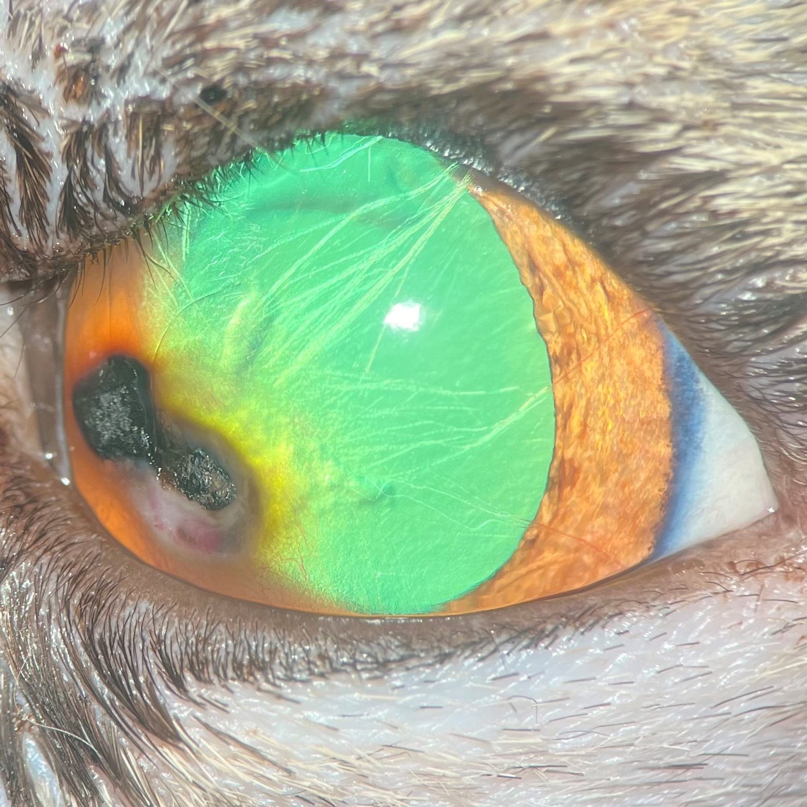
Corneal Sequestrum Treatment for Cats
A corneal sequestrum is a brown to black area of dead tissue on the cornea, the clear front surface of the eye. It is seen almost exclusively in cats and can be painful and slow to resolve. At Animal Eye Clinic, Dr. Robert M. Gwin diagnoses corneal sequestrum and performs the delicate corneal surgery often needed to remove it, at our Tulsa and Oklahoma City offices.
Request an AppointmentWhat Is a Corneal Sequestrum?
A corneal sequestrum, sometimes called corneal necrosis or corneal mummification, develops when an area of the cornea dies and becomes discolored. It usually starts as a faint amber or brown spot in the center of the cornea and can darken to black as it progresses. The body often reacts to the dead tissue with inflammation and blood vessel growth around the lesion.
Causes and Risk Factors
The exact cause is not fully understood, but corneal sequestrum is commonly associated with:
- Chronic corneal irritation or non-healing corneal ulcers
- Feline herpesvirus infection
- Flat-faced breeds such as Persians, Himalayans, and Burmese
- Prominent eyes, incomplete blinking, or entropion
- Tear film problems or dry eye
Signs of Corneal Sequestrum
Cats with a sequestrum may have a visible brown or black spot on the eye, squinting, tearing, discharge, redness, and a tendency to keep the eye partially closed. Some cats show surprisingly little discomfort, while others are quite painful.
Corneal Sequestrum Treatment
Treatment depends on the size and depth of the sequestrum and how uncomfortable your cat is. Small, superficial lesions in comfortable cats are sometimes managed medically with lubricating and antiviral or antibiotic medications while the body attempts to slough the lesion, though this can take weeks to months.
In many cases, surgical removal is the best option. Dr. Gwin performs a keratectomy under an operating microscope to remove the diseased corneal tissue. If the removal leaves a deep defect, a grafting procedure such as a conjunctival flap may be placed to strengthen the cornea and support healing. Addressing underlying factors such as herpesvirus or eyelid problems helps reduce the risk of recurrence. Learn more about our surgical procedures and other ophthalmic diseases we treat.
Corneal Sequestrum FAQs
What does a corneal sequestrum look like?
It appears as an amber, brown, or black spot on the clear surface of the cat’s eye, often with surrounding redness or blood vessels.
Which cats get corneal sequestrum?
Flat-faced breeds such as Persians, Himalayans, and Burmese are most at risk, but any cat with chronic corneal irritation or feline herpesvirus can develop one.
Is corneal sequestrum painful?
It can be. Many cats squint, tear, and keep the eye partly closed, though some show only mild signs.
Will a corneal sequestrum go away on its own?
Some small lesions slough off over weeks to months, but many require surgical removal to resolve pain and prevent complications.
How is corneal sequestrum treated surgically?
Dr. Gwin removes the diseased tissue with a keratectomy and may place a conjunctival flap graft if the defect is deep.
Can a corneal sequestrum come back?
Recurrence is possible, especially if underlying factors like feline herpesvirus or eyelid problems aren’t managed.
Schedule an Exam for Your Cat
If you notice a brown or black spot on your cat’s eye, call (918) 632-0508 (Tulsa) or (405) 751-3821 (Oklahoma City) to schedule an evaluation with Dr. Gwin.