

Corneal Dystrophy Treatment for Dogs and Cats
Dystrophy is the degeneration of tissues due to disease or malnutrition, and it is frequently inherited. As with all diseases of the eye, corneal dystrophies vary in cause and severity, so treatment is very specific to your pet’s condition. At Animal Eye Clinic, Dr. Robert M. Gwin diagnoses and treats superficial corneal dystrophy and corneal endothelial cell dystrophy at our Tulsa and Oklahoma City offices.
Request an AppointmentSuperficial Corneal Dystrophy
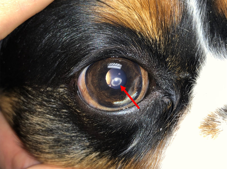
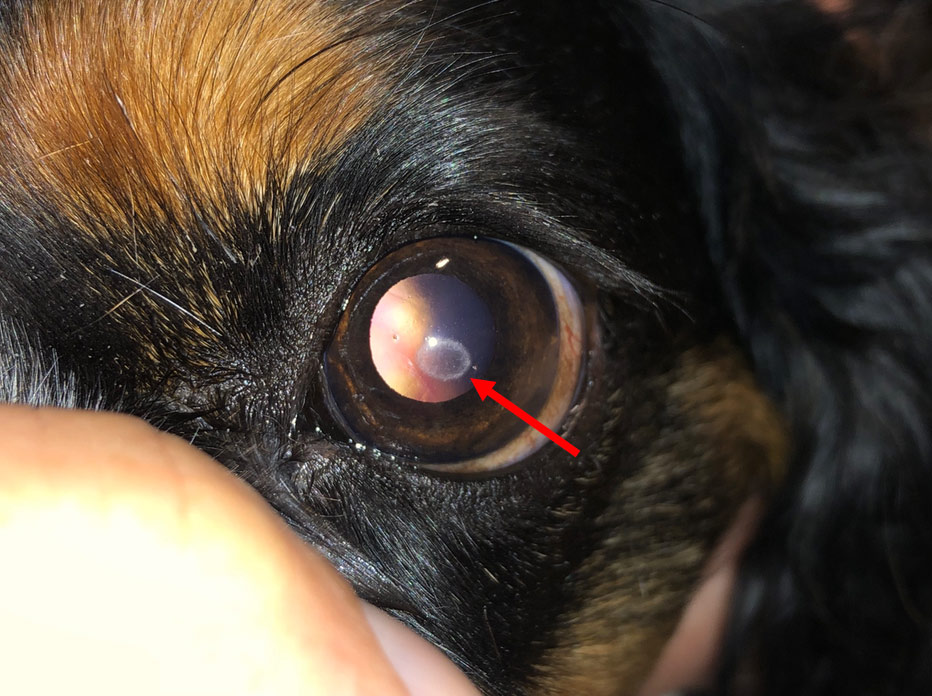
Superficial corneal dystrophy is characterized by white, opaque mineral or lipid deposits, such as cholesterol or calcium, within the cornea, the clear front part of the eye. The size, shape, and density of these plaque-like deposits vary widely and can cause painful inflammation (keratitis) and secondary corneal ulcers. Although the affected areas can be very visible, they rarely cause blindness, and more severe cases may require surgery.
Treatment for Superficial Corneal Dystrophy
Our goal in treating superficial corneal dystrophy and corneal degeneration with topical medication is to improve the health of the normal cells covering the corneal surface.
In cases of severe progression and discomfort, much of the mineral can be removed through a superficial keratectomy, a surgery that removes the outer layer of the cornea along with the deposits. If a deep ulcer develops as a result of the degeneration, a grafting procedure called a conjunctival flap may be necessary to help the cornea heal. Learn more about our surgical procedures.
Corneal Endothelial Cell Dystrophy and Degeneration
The cornea is normally the transparent “windshield” of the eye, protecting the internal structures while allowing light to enter. The endothelium is a single layer of cells lining the inside of the cornea. Though only one cell thick, this layer is vital for keeping the cornea clear and maintaining vision. The cornea stays clear thanks to a very regular arrangement of supportive stromal fibers kept in a relatively dehydrated state. If water builds up, that arrangement is disrupted and the cornea loses its clarity.
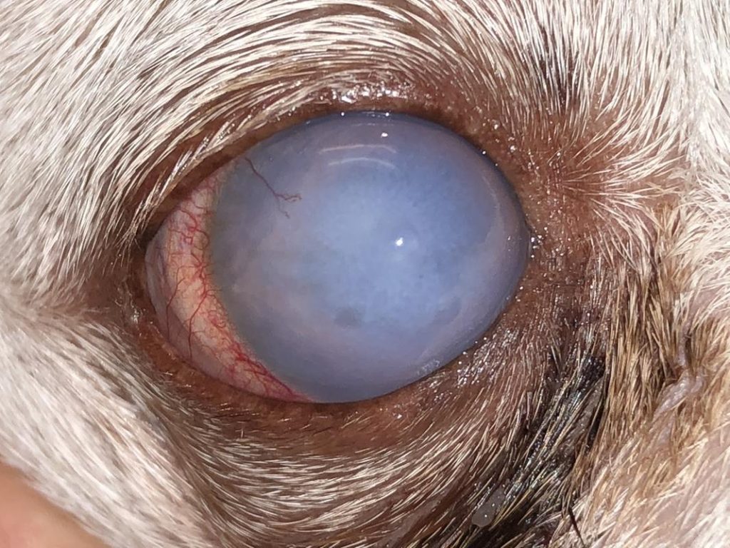
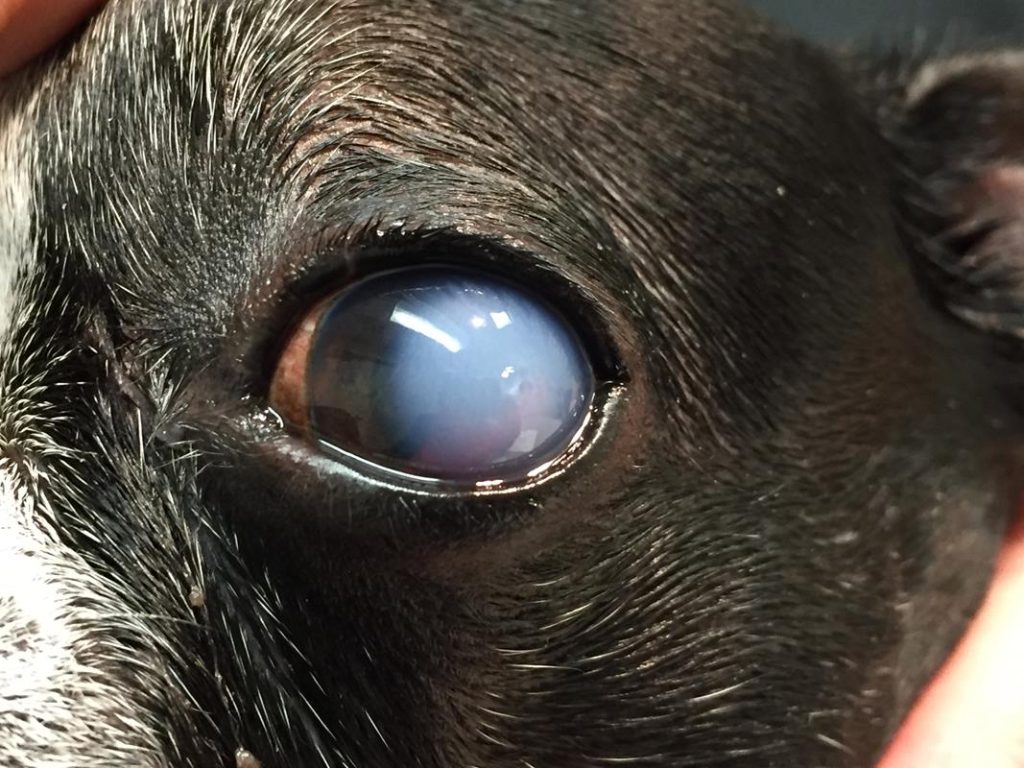
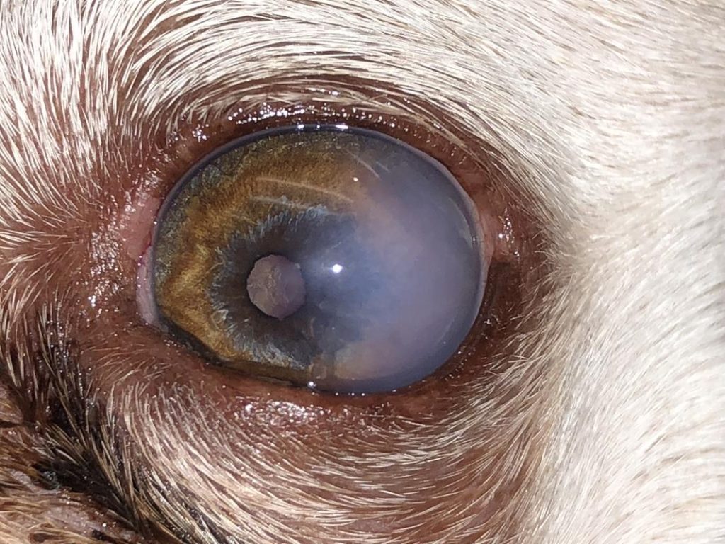
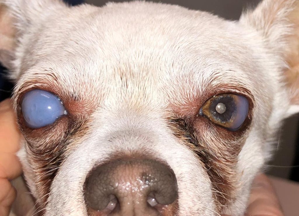
Endothelial cells regenerate very slowly and incompletely. When cells are lost, the remaining cells spread out to cover the gap. As long as the number of functioning cells stays above a critical threshold, they keep the cornea clear. If the number falls below that threshold, the cornea can no longer stay dehydrated, and a progressive blue cloudiness called corneal edema develops.
This can occur as an inherited condition in certain breeds, such as Dachshunds, Chihuahuas, and Boston Terriers, where it is called endothelial cell dystrophy. Endothelial degeneration can also occur secondary to many other eye disorders, including uveitis, elevated eye pressure from glaucoma, lens luxation, and endothelialitis, which is often thought to be immune mediated.
Fluid in the cornea is not painful by itself, but it can cause small blisters called bullae. If these bullae reach the surface and rupture, they leave a painful corneal ulcer. These ulcers can heal quickly with appropriate medication, but if they become a recurring problem, a surgical procedure may be recommended to minimize bullae formation and help the erosion heal.
Treatment for Corneal Endothelial Cell Dystrophy
This condition can be stabilized, and in some cases partially reversed, with proper medical therapy. Medications that can help include judicious use of topical corticosteroids, NSAIDs, immunosuppressive drops, and antibiotic drops, especially if a corneal ulcer occurs.
Surgical therapy is usually aimed at the complications of endothelial dystrophy, such as bullae that rupture into ulcers. A bandage contact lens and a temporary partial eyelid closure (temporary tarsorrhaphy) can protect and support the corneal surface while it heals.
Corneal Dystrophy FAQs
What does corneal dystrophy look like in dogs?
Superficial dystrophy appears as white or gray crystalline spots on the cornea, while endothelial dystrophy causes a gradual blue cloudiness across the eye.
Is corneal dystrophy painful?
The deposits or fluid alone are often not painful, but inflammation, ulcers, or ruptured bullae can cause significant discomfort.
Which breeds are prone to endothelial dystrophy?
Dachshunds, Chihuahuas, and Boston Terriers are among the breeds known to inherit corneal endothelial cell dystrophy.
Will my pet go blind from corneal dystrophy?
Superficial deposits rarely cause blindness. Endothelial dystrophy can reduce vision as corneal edema progresses, but medical therapy can often stabilize it.
Can corneal dystrophy be cured?
It usually can’t be cured, but it can often be managed and stabilized with medication, and surgery can address deposits or recurrent ulcers.
What surgery is used for corneal dystrophy?
Options include superficial keratectomy to remove deposits, conjunctival flap grafting for deep ulcers, and a bandage contact lens with temporary tarsorrhaphy to protect the cornea.
Concerned About Cloudiness in Your Pet’s Eye?
Call (918) 632-0508 (Tulsa) or (405) 751-3821 (Oklahoma City) to schedule an exam with Dr. Gwin.